How Do You Diagnose a Virus Infection in a Plant?
By a combination of the following ...
1.) Inclusions
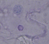
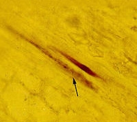
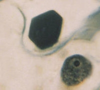
Staining properties: There are two stains used in looking for viral inclusions. One is Azure A (AA), which stains RNA pink (Figure 1 and 2) and DNA blue. The second stain is the orange-green (OG) stain (Figure 3), which stains protein.
 (Figure 1) |
 (Figure 2) |
 (Figure 3) | original (Figure 4) |
Some inclusions, such as the cylindrical inclusions of most potyviruses stain only in OG. Others like cucumber mosaic displayed in Figure 1 will stain with both stains. Tobacco mosaic inclusions like the one in Figure 3 will stain with both stains, but it will only stain in AA when it's heated. Differential staining can help in diagnosis.
Location: Many inclusions are found in the epidermis (Figure 1 and 3), but others can only be found in the vascular tissue like Citrus tristeza virus displayed in Figure 2. Those of Geminiviruses and Rhabdoviruses (Figure 4) are found in the nucleus. The location of the inclusion also helps to narrow down the type of virus infecting the plant.
2.) Host Range Inoculations
Over the years plant virologists have inoculated plant viruses to many different plants. Each known plant virus has a list of susceptible plants and insusceptible plants.
By grinding a sample of an infected plant in a buffer (Figure 5), you can manually inoculate other plants (Figure 6). Those that become infected can be compared with the list of plants for a suspected virus.
 (Figure 5) |
 (Figure 6) |
If the host range of your unknown virus is the same as that of some known virus you have another clue as to what virus you may have.
3.) Electron Microscopy
Virus particles can be seen only with the use of a very powerful microscope called an electron microscope (EM). Plant sap containing virus particles can be prepared and put into an electron microscope and examined for virus particles. There are several basic shapes that virus particles take. By viewing the particle in the EM you can narrow down the type of virus you have.
Four different shapes are shown in Figure 7-10. A rhabdovirus will have a bullet-like shape (Figure 7). Tobamoviruses like tobacco mosaic have rigid rods (Figure 8). Potyviruses (Figure 9) and clostroviruses have flexous rods. Many other viruses have a spherical shape (Figure 10).
original (Figure 7) original (Figure 8) original (Figure 9) original (Figure 10) |
 (Figure 11a) 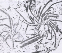 (Figure 11b) 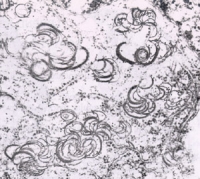 (Figure 11c) |
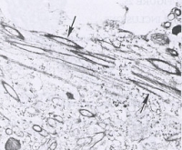
Plant tissue can also be fixed, thinly sectioned, and viewed under the EM. If you were to see something like that on the right you would know you had a potyvirus.
4.) Serological Tests
Viruses can be purified and the purified virus injected into a mammal such as a rabbit. The inoculated animal will make antibodies to protein coat of this virus. The animal is bled and the serum (antiserum) that results can used to detect plant viruses. Antibodies can also be made with molecular techniques.
One type of serological test is called immunodiffusion. In the picture below (Fig. 12) the antiserum was put into the middle well. The two wells on the top and the middle left in Fig. 12 contain a virus identical to the virus used to make the antiserum. The two wells shown in top and middle right of Fig. 12 are a related virus. The two bottom wells are healthy controls.
 (Figure 12) |
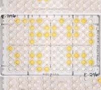 (Figure 13) |
The picture in Figure 13 is an example of test called an ELISA test. There are several different procedures for this type of test but the end results are the same. The wells that are yellow are positive for the virus (they reacted with the antiserum). The other wells are either healthy controls or negative for the virus.
5.) DNA Tests
Tests can also be done that will detect the nucleic acid of the virus.
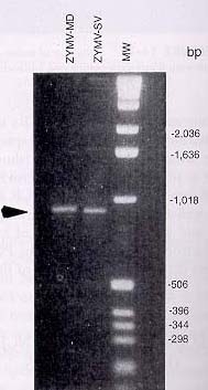
An example of a potyvirus detected in a sample by PCR (polymerase chain reaction) is displayed in Figure 14.
A test using a DNA probe which attaches to the sample when the virus is present is displayed in Figure 15. The dark spots are positive for the virus and those that do not react are negative.
 (Figure 14) |
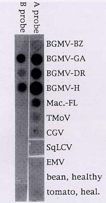 (Figure 15) |
There is another type of test which can detect the double stranded form or replicating form of some RNA viruses. Using appropriate controls, the size of the band on a gel can tell you what type of virus you have. PCR Products can also be sequenced and compared to sequences in the GenBank.